


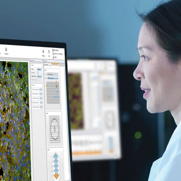
Cellular Imaging Software
Acquire, visualize, analyze, discover
Revvity offers sophisticated cellular imaging and analysis software to help you acquire, visualize, analyze and share your image data more effectively and efficiently, so you can spend more time discovering.
From image cytometry, to live-cell imaging and sophisticated sub-cellular high-content imaging and analysis, our cell image analysis software is workflow-driven, guiding you through the process step-by-step. In addition, we provide software systems to help you efficiently manage the terabytes of scientific imaging data that can be generated by advanced high-content screening experiments.
For research use only. Not for use in diagnostic procedures.


Filters
1 - 25 of 28 Resources
3D cell culture workflow solutions
More than ever, researchers are turning to 3D cell cultures, microtissues and organoids to bridge the gap between 2D cell cultures and in vivo animal models. Our solutions-based approach is designed to support you at every step of the cell culture workflow so you can seamlessly culture, treat, and analyze 3D cell cultures. Begin generating more physiologically relevant data to better understand biological processes and make informed decisions with our solutions. For research use only. Not for use in diagnostic procedures
Analysis of mitochondrial dynamics in human iPSC-derived neurons using the Operetta CLS High-Content Analysis System
Mitochondrial dynamics are essential for energy conversion and neuron survival. Understanding changes in mitochondrial dynamics is therefore crucial in developing mitochondria-based therapy options for complex pathological conditions such as cancer, neurological disorders, and metabolic syndromes. Fast kinetic live-cell imaging combined with high-content screening represents a promising strategy to quantitatively track these changes in real time and at large scale. This application note demonstrates how to investigate mitochondrial dynamics in human iPSC-derived neurons using the Operetta ® CLS ™ high-content analysis system. Fast frame rate imaging to accurately capture rapid cellular responses Reliable quantification of mitochondrial dynamics in neurons Flexible kinetic acquisition settings for each channel to avoid unnecessary data acquisition
Analyzing ERK signal transduction in live cells using a FRET-based biosensor
Extracellular signal-regulated kinase (ERK) is a key component in the regulation of embryogenesis, cell differentiation, cell proliferation, and cell death. The ERK signaling pathway is altered in various cancer types and is frequently investigated as a target for therapeutic intervention. This application note describes how a live cell FRET assay to study ERK signaling was performed on the Operetta CLS ™ high-content analysis system. The optimized design of the FRET-based biosensor, the high-quality imaging of the Operetta CLS system and the easy-to-use image analysis tools of the Harmony™ software contribute to the robustness of the high-content assay.
Artificial intelligence, machine learning and deep learning: applications in cellular imaging for improved drug discovery productivity
There has been a lot of buzz around artificial intelligence, machine learning and deep learning. Is the reality living up to the hype? In the world of cellular imaging and its application to drug discovery, there is evidence of real progress against some of the critical challenges facing scientists using these technologies. In this white paper, you will learn about: Challenges in cellular imaging and drug discovery that Artificial Intelligence (AI), Machine Learning (ML) and Deep Learning (DL) are helping to overcome How these technologies are used by leading cellular imaging scientists An outlook to how AI, ML and DL in cellular imaging have the potential to further advance drug discovery and improve productivity in the future
Cell painting: your questions answered
With powerful potential for phenotypic drug discovery, cell painting is being rapidly adopted by leading scientists across the globe. However, implementing cell painting assays can present some challenges. To help you avoid some of the common pitfalls, we’ve collated answers from our experts to the top questions that we’ve been hearing from scientists and researchers. You will learn: How to choose the cell model What to look for in labelling reagents Key considerations for image acquisition Recommendations for image analysis Data analysis approaches
Cytotoxicity studies on 3D primary liver microtissues
Liver toxicity remains one of the main reasons for drug failure in clinical trials. Improving preclinical toxicity testing is a major prerequisite for improving the drug de-risking process. Early compound de-risking decisions require in vitro data with the highest possible biological relevance. Three dimensional (3D) microtissues, or spheroids, are one of the most well characterized models for 3D culture and cell-based drug screening due to their reproducibility and similarity to the in vivo situation.
Deeper insights from your 3D cell model imaging
Researchers are increasingly looking to 3D cell models to bridge the translational gap between 2D cell cultures and in vivo conditions. These cell models more closely represent the microenvironments, cell-to-cell interactions, and biological processes that occur in vivo. Download our infographic to discover tools and strategies to get the most out of your 3D cell model imaging and analysis workflows.
Distinguishing cell types by phenotypic profiling of the nucleus
The promise of high-content screening is the acceleration of discovery by extracting as much relevant information as possible from cells. Nevertheless, a large percentage of high-content screens analyze only a small number of image-based properties. As a result, valuable information from precious cells and disease models is not utilized. As nearly all screening approaches require a nuclear counterstain such as Hoechst to facilitate segmentation, phenotypic profiling of the nuclei can offer new and additional perspectives on assays at no extra cost.
Fast Kinetic Calcium Flux Imaging Using the Opera Phenix Plus High-Content Screening System
Dysregulation of GPCR signaling, particularly calcium signaling, has been implicated in different diseases, e.g. cancer and Alzheimer’s disease. Currently about 34% of all FDA approved drugs target GPCRs indicating their importance for drug discovery, and in vitro cellular GPCR assays are important tools to identify new drugs. Our application note describes how to analyze fast changes in intracellular calcium levels upon GPCR stimulation and inhibition using a fluorescence imaging assay on the Opera Phenix® Plus high-content screening system. Measure fast kinetic assays using the onboard liquid handling module with tip-based pipettor Track changes with high temporal resolution through acquisition at frame rates of up to 100 frames per second Analyze agonist- and antagonist-dependent changes in calcium concentration with single cell resolution
Harness the power of 3D cell model imaging
3D cell model imaging and analysis workflows provide more physiologically relevant insights but are not without their challenges. Whether you’re familiar with high-content screening or you want to take your analysis of 3D cell models to the next level, you’ll need the right tools and strategies to overcome them. This guide provides hints and tips for successful 3D cell model imaging. Learn how to: Rapidly generate high-quality images from large, thick cell samples with confidence Reduce imaging times and handle the increased volumes of data Seamlessly analyze your images without tedious transfer between software
HCS featured publications for infectious disease
This provides a list of various HCS publications in the area of infectious disease (viral, parasitic, and bacterial).
HCS Publication List
A selection of publications from 2022 and 2023 using Revvity HCS systems are featured here and categorized by research area.
High content screening in three dimensions
Researchers are increasingly looking to 3D cell cultures, microtissues, and organoids to bridge the gap between 2D cell cultures and in vivo animal models. This whitepaper documents a streamlined procedure for getting the most information, as quickly as possible, using solutions from Revvity.
High-content imaging of brain-on-chip microfluidic devices using PreciScan intelligent acquisition.
Brain-on-chips (BoC) are innovative tools that simulate complex brain environments, enhancing our understanding of neurodegenerative diseases (NDDs). While a range of BoC platforms have been developed to model diverse NDD pathophysiologies, the various channel architectures of these microfluidic devices present challenges in image acquisition and analysis. High-content imaging protocols must be adaptable to these differing designs. This technical note, developed in collaboration with ETAP-Lab, demonstrates an automated image acquisition protocol for BoC microfluidic devices. The protocol utilizes the Operetta CLSTM high-content analysis system with PreciScan intelligent acquisition to effectively image neuronal cells in microfluidic devices. We address: - How to automate image acquisition of microfluidic devices - How to set up a pre-scan re-scan routine for selected relevant channels within brain-on-chip devices - How to increase throughput for quantitative microscopy-based screening of brain-on-chip models
How to perform long term live cell imaging in a high‑content analysis system
Live cell imaging has gained importance within drug discovery over recent times, as researchers look for more meaningful insights into cellular behavior and function. However, setting up live cell experiments can be challenging, as temperature, CO 2 and evaporation need to be controlled to ensure optimal cell growth conditions. In this technical note, we demonstrate: Successful five day live cell imaging on Operetta CLS and Opera Phenix high-content systems Avoidance of phototoxicity with gentle digital phase contrast imaging Analysis of cell growth and morphology on a single cell level without any fluorescence staining.
How to perform successful long term live cell imaging in a high-content analysis system
Live cell imaging has gained importance within drug discovery over recent times, as researchers look for more meaningful insights into cellular behavior and function. However, setting up live cell experiments can be challenging, as temperature, CO2 and evaporation need to be controlled to ensure optimal cell growth conditions. In this technical note, we demonstrate: Successful five-day live cell imaging on Operetta CLS and Opera Phenix high-content systems Avoidance of phototoxicity with gentle digital phase contrast imaging Analysis of cell growth and morphology on a single cell level without any fluorescence staining
Imaging bile canaliculi in 3D liver microtissues
Analyzing transport of biliary metabolites is essential to predict pharmacokinetics and hepatotoxicity during drug development. A functional impairment of hepatobiliary transporters, such as bile salt export pump (BSEP) and multidrug resistance-associated protein 2 (MRP-2), is strongly associated with an increased risk of cholestatic liver injury. Here, we describe a 3D high-content screening assay to study hepatobiliary transporter function in InSphero ™ human liver microtissues. Confocal imaging and automated image analysis were used to quantify BSEP and MRP-2-mediated efflux of fluorescent substrates into bile canaliculi.
Label-free analysis of cardiomyocyte beating using the Opera Phenix Plus System
Drug-induced cardiotoxicity is a frequent cause of drug attrition during drug development and can lead to the withdrawal of drugs from the market, so non-invasive approaches that allow quick and reliable assessment of the cardiotoxic potential of a compound are key to improve drug safety. In this application note, learn how the Opera Phenix ® Plus high-content screening system together with the MuviCyte ™ live-cell imaging system can be used for: Reliable quantification of cardiomyocyte beating frequency Fast frame rate imaging up to 100 fps to accurately capture fast cellular responses Tip-based on-board pipetting compatible with 96 and 384-well plates Stable environmental conditions with temperature, CO2, and humidity control
Measuring FRET using the Opera™ Phenix™ high-content screening system
Fundamental processes in living cells, such as apoptosis and signal transduction are controlled by proteins, often acting in concert with other protein partners through protein-protein interactions (PPIs). Inappropriate protein-protein recognition can fundamentally contribute to many diseases, including cancer. Therefore, inhibiting protein-protein interactions represents an emerging area in drug design.
Nuclei segmentation on brightfield images using a pre-trained Artificial Intelligence (AI) model
The use of Artificial Intelligence (AI) for analysis of cellular images is very promising, especially for image segmentation tasks that were previously challenging or unfeasible, such as the segmentation of nuclei on brightfield images. Brightfield images, while being widely accessible, often suffer from low contrast and lack the specific staining that fluorescent images provide. Download our technical note to learn more about Phenologic.AI: Robust segmentation of cells on brightfield images Ready-to-use pre-trained AI model compatible with different magnifications and cell lines Digital phase image reconstruction for cytoplasm segmentation For research use only. Not for use in diagnostic procedures.
Opera Phenix High-Content Screening System Technical Performance: Speed
The Opera Phenix™ high-content screening system’s state of the art hardware, combined with Harmony® imaging and analysis software, improves acquisition and analysis speed, enabling users to perform highly multi-parametric phenotypic screens effectively. In this technical note, learn how the Opera Phenix system’s Synchrony™ Optics and sCMOS cameras image up to 6x faster, how water immersion objectives combined with binning can reduce exposure time by approx. 20-fold and how image acquisition and analysis time can be reduced by > 70% compared to a classical spinning disc confocal system.
Opera Phenix Plus High-Content Screening System Brochure
The Opera Phenix® Plus High-Content Screening System is the premier confocal solution for today's most demanding high content applications.
Operetta CLS High-Content Analysis System Brochure
The product of more than a decade of experience in high-content screening, the Operetta CLS delivers the speed and sensitivity for everyday assays, and even complex challenges – live cells, phenotyping, rare events, and much more.
Phenotypic analysis of CRISPR-Cas9 cell-cycle knockouts using cell painting
Combining CRISPR gene editing and cell painting provides researchers with the ability to correlate genotypes with phenotypes and holds the promise to help understand gene functions in more detail and at greater scale. This application note demonstrates a harmonized workflow for morphological profiling of CRISPR-Cas9-mediated knockouts using cell painting, with simplified feature extraction using the dedicated Cell Painting Building Block in Harmony ® High-Content Analysis software. Workflow for an arrayed CRISPR knockout screen using Cas9 stable cells and Edit-R ® synthetic single guide RNAs Phenotypic analysis of gene knockouts with the PhenoVue ™ Cell Painting JUMP kit and the Opera Phenix ® Plus High-Content Screening system One step solution for cell painting feature extraction in Harmony High-Content Analysis software Phenotypic clustering using Principal Component Analysis and Self-Organizing Maps
Phenotypic analysis of hypertrophy in human iPSC-derived cardiomyocytes
Discover how a phenotypic screening assay to study time-dependent effects of endothelin-1-induced hypertrophy was set up using human induced pluripotent stem cell (hiPSC)-derived cardiomyocytes. Learn how to: Differentiate early and late effects in endothelin-1-induced hypertrophic cardiomyocytes Leverage robust and fast analysis approaches without single cell segmentation Analyze phenotypic changes of cardiac hypertrophy with SER and Haralick texture features