Total list price:
Log in to view your online price USD 408.57
Select your location.
*e-commerce not available for this region.


 View All


SAVE 10% at Revvity.com or through your punchout with promo code.
SEPT25
Ends Sept. 23 - Terms and conditions apply.
| Feature | Specification |
|---|---|
| Color | Red |
| Filter | Cy5 |
| Organelle and Cell Compartment | Mitochondria |
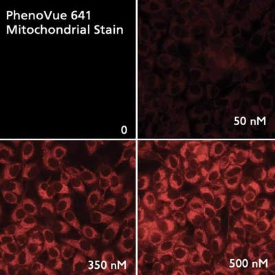
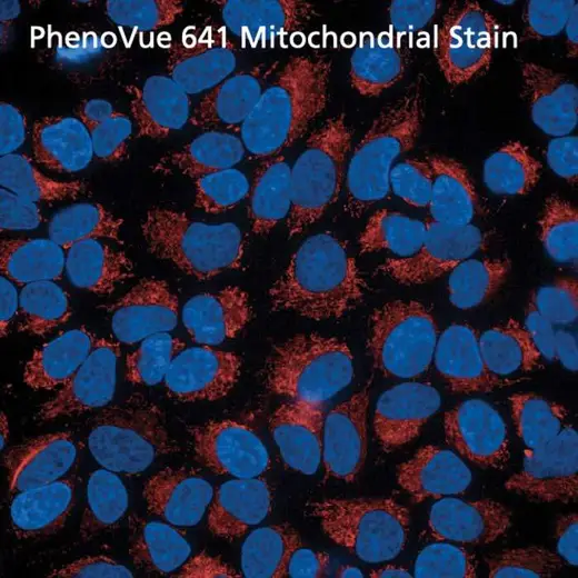
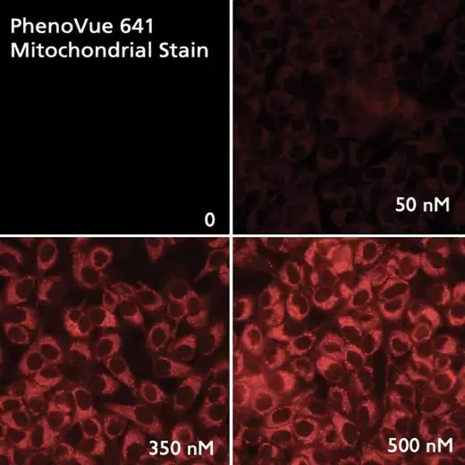
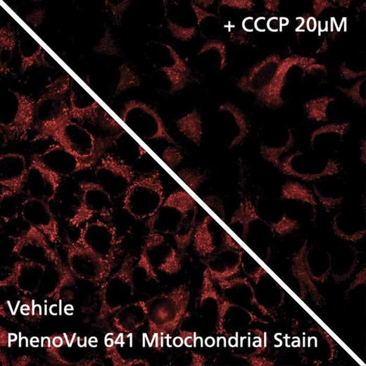
Fluorescent mitochondrial stains are commonly used for visualizing and quantifying mitochondria whose dysfunctions, including impaired biogenesis, dynamics or trafficking, are a hallmark of some human diseases, such as neurodegenerative disorders.
PhenoVue 641 Mitochondrial stain can be used to detect mitochondria in indirect immunofluorescence, immunohistochemistry and flow cytometry, as well as high-content analysis and screening applications.
Typical working concentration: 100 nM (53 ng/mL)
Equivalent number of microplates:
| Color |
Red
|
|---|---|
| Form |
Desiccated
|
| Maximum Emission Wavelength (Emmax) |
662 nm
|
| Maximum Excitation Wavelength (Exmax) |
641 nm
|
| Application |
High Content Imaging
Microscopy
|
|---|---|
| Brand |
PhenoVue™
|
| Detection Modality |
Fluorescence
|
| Filter |
Cy5
|
| Organelle and Cell Compartment |
Mitochondria
|
| Quantity |
20 x 50 µg (92 nmoles)
|
| Sample Type |
Live cells with fixation post staining
|
| Shipping Conditions |
Shipped in Dry Ice
|
| Storage Conditions |
-16 °C or below, protected from light
|
| Type |
Individual reagent
|
Are you looking for resources, click on the resource type to explore further.
This is a product information sheet for PhenoVue Mitochondrial Stains.
Are you looking for technical documents related to the product? We have categorized them in dedicated sections below. Explore now or request your COA/TDS, SDS, or IFU/manual.


")


We are here to answer your questions.


.")